Cnr Eloff & Albertina Sisulu, Johannesburg, RSA
+2711 333 8160
mreyesoptometrist@gmail.com
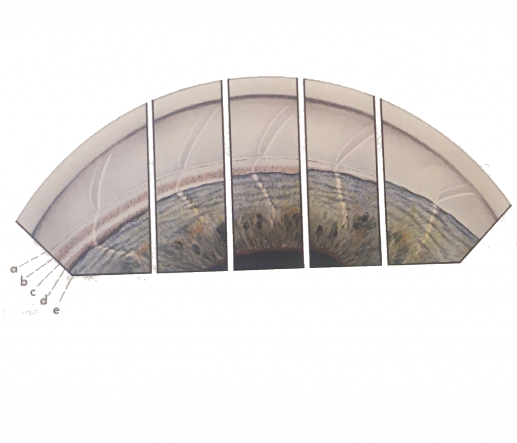
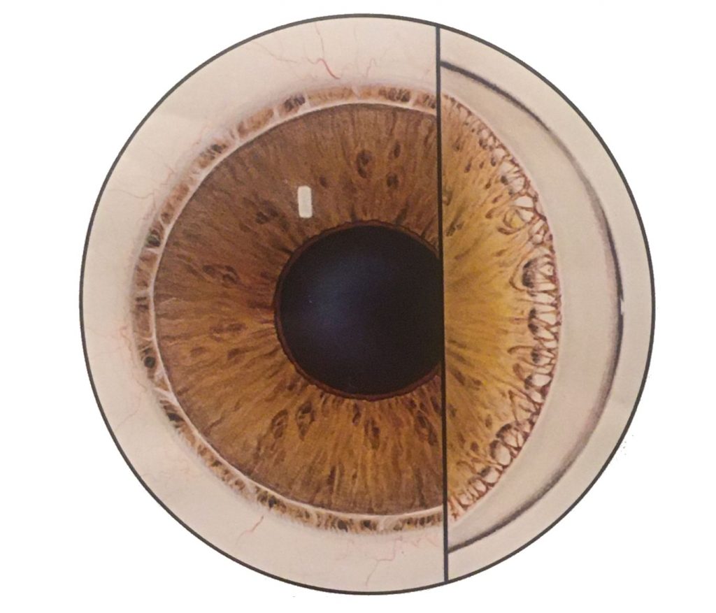
Various widths of the normal angle seen in consecutive inserts. Note that the corneal optical wedge narrows as it approaches Schwalbe’s line and seems to end there in the situation of a narrow angle. a) Schwalbe’s line; b) Trabecular meshwork; c) Scleral spur; d) Ciliary body band; and e) Peripheral iris.
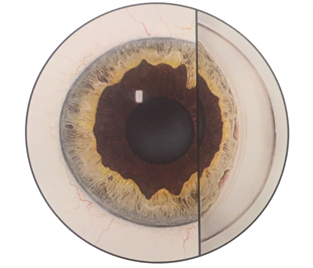
Prominent congenital ectropion uveae involving much of the iris surface. Gonioscopy reveals irregular insertion of the iris into the angle.

Frontal view showing inferiorly dislocated lens in a patient with homocystinuria. Upper left hand insert showing dislocated lens with normal zonular attachments as seen in Marfan syndrome. Upper right hand insert shows dislocated lens with defective zonular apparatus as seen in the homocystinuria.
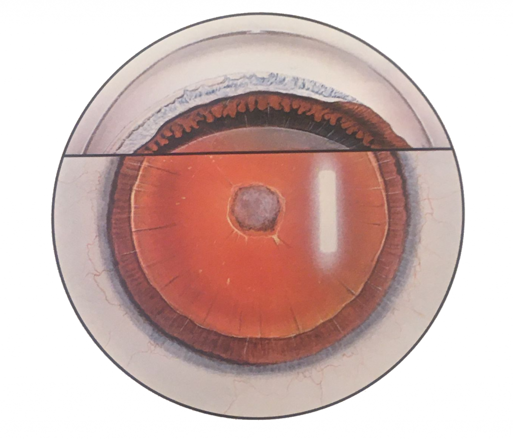
Slit-lamp view showing the absence of iris and typical anterior polar cataract. Gonioscopic view showing residual iris stump, peripheral anterior synechiae, and apposition of the iris to the trabecular meshwork exposing the pigment epithelium.

Slit beam showing shallow anterior chamber. Lower right insert shows gonioscopic appearance of a narrow angle. Upper rigt insert shows the same angle with indentation gonioscopy demonstrating typical peripheral anterior synechiae to the trabecular meshwork.

The anterior segment including the angle appears to be within normal limits.
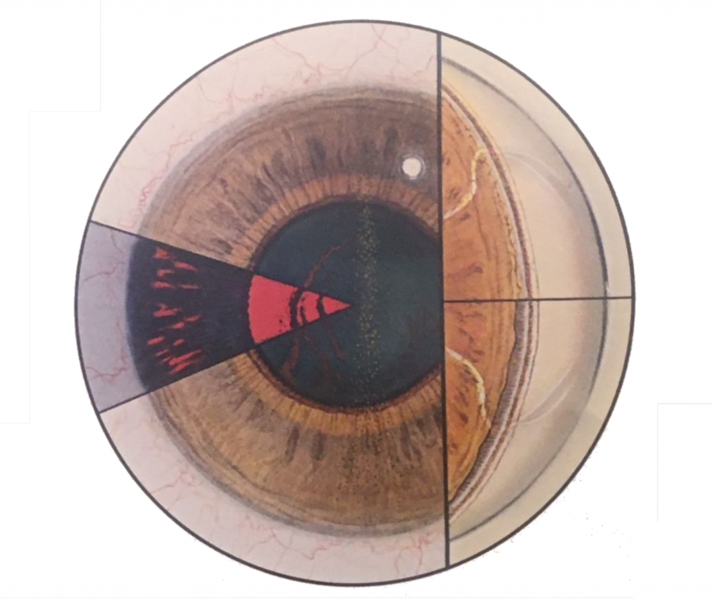
Frontal view showing the depositionof pigment on the endothelial surface of the cornea (Kruckenberg spindle) and on the posterior surface of the lens. Left insert shows typical radial spokelike translluminatory defects in the mid-peripheral iris as well as pigmentation of the posterior lens surface. Right insert (below) shows gonioscopic appearance of lower angle with dense…
Read more
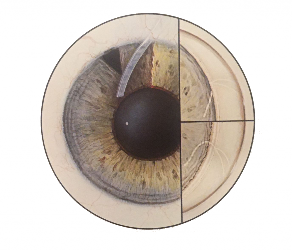
On direct view, a prominent Schwalbe’s line can be seen to encircle the peripheral cornea. Strands of peripheral iris tissue attach to Schwalbe’s line and are obvious in the gonioscopic view.
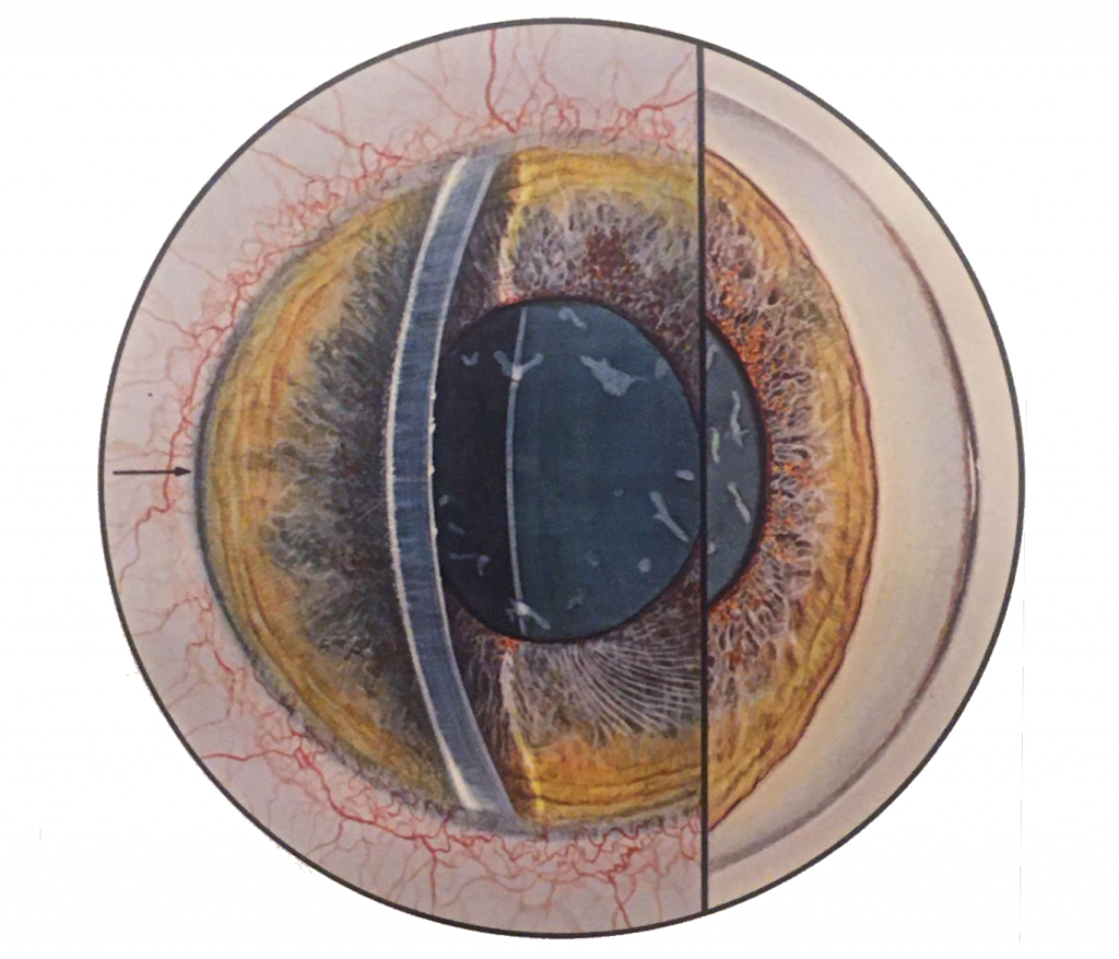
Axial portion of the anterior chamber is of normal depth and an iridectomy is present at 11 o’clock. The plane of iris is vertical, and the iris root undergoes a knee-shaped bend (insert lower right) in the extreme periphery, cauing a sudden narrowing of the angle in the presence of a patient iridectomy. Insert upper…
Read more
Frontal view showing shallow anterior chamber, diffuse iris stromal atrophy, spiraling of the iris stroma inferiorly, and glaukomflecken of the lens. Gonioscopy shows permanent synechial closure.