
Pigmentary Glaucoma
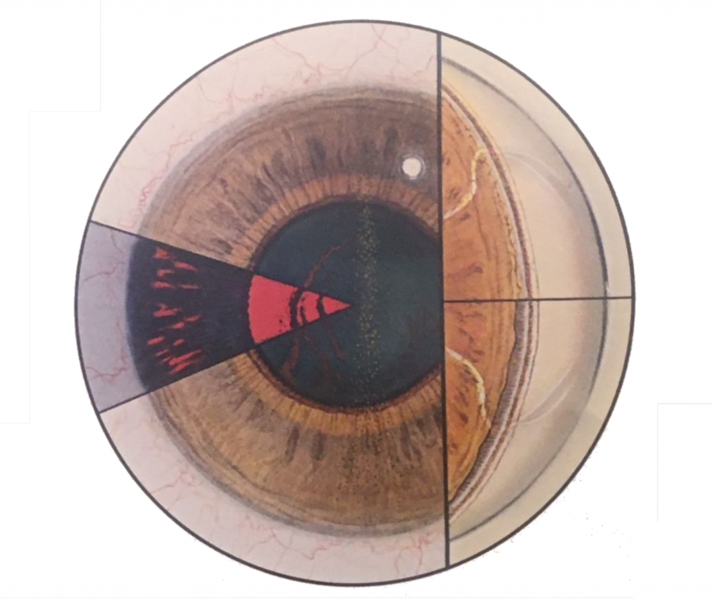
Frontal view showing the depositionof pigment on the endothelial surface of the cornea (Kruckenberg spindle) and on the posterior surface of the lens. Left insert shows typical radial spokelike translluminatory defects in the mid-peripheral iris as well as pigmentation of the posterior lens surface. Right insert (below) shows gonioscopic appearance of lower angle with dense homogenous pigment in the posterior trabecular meshwork and a concave appearance to the peripheral iris with a wide ciliary body band. Right insert (above) shows an upper angle with similar pigmentation and narrow ciliary body band.